A team of researchers has developed a new tool to probe the composition of our cells with much more precision. Ultimately, this pilot study could help to better understand the molecular basis of many diseases.
Many human diseases are the result of malfunctions in our cells. A tumor, for example, is able to grow because a gene has not been precisely translated into a particular protein. To try to better combat these dysfunctions, scientists must therefore understand all the components of our cells . However, this is not the case today.
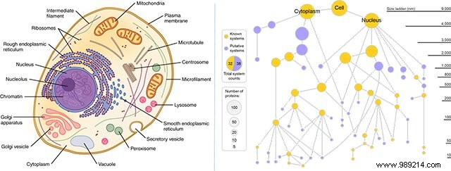
"If you imagine a cell, you're probably imagining the colored diagram from your cell biology textbook, with the mitochondria, endoplasmic reticulum, and nucleus. But is that the whole story? Definitely not “, details Dr. Trey Ideker, of the San Diego School of Medicine.
Scientists have long realized that our cells are much more structurally complex than we think , but until now, much of this information was inaccessible to us. In this sense, progress is being made.
By combining microscopy, biochemical techniques and artificial intelligence, researchers at the University of California School of Medicine have indeed recently taken a big step in forward in understanding human cells. The technique, known as Multi-Scale Integrated Cell (MuSIC), has just been the subject of a study published in Nature.
Some microscopes allow scientists to see down to the level of a single micron, revealing certain organelles such as mitochondria. On the other hand, biochemical techniques allow scientists to go down to the nanometric scale. Here, an artificial intelligence program has made it possible to bridge the gap between the nanometer scale and the micron scale .
As part of this work, MuSIC has revealed about seventy components contained in a human kidney cell line, half of which was unknown . These were mostly proteins that bind to RNA. These protein complexes, previously invisible to us, are thought to be involved in the splicing that allows genes to be translated into proteins and helps determine which genes are activated when.

Note that system does not yet map the content of cells at specific locations, as is the case conventionally, partly because their locations are not necessarily fixed , evolving according to the type of cell and the situation.
Finally, remember that this study only focuses on a single type of cell. Now, the researchers plan to build on this tool to analyze different cell types from different species. Ultimately, we could then better understand the molecular basis of many diseases by comparing what is different between healthy and diseased cells.